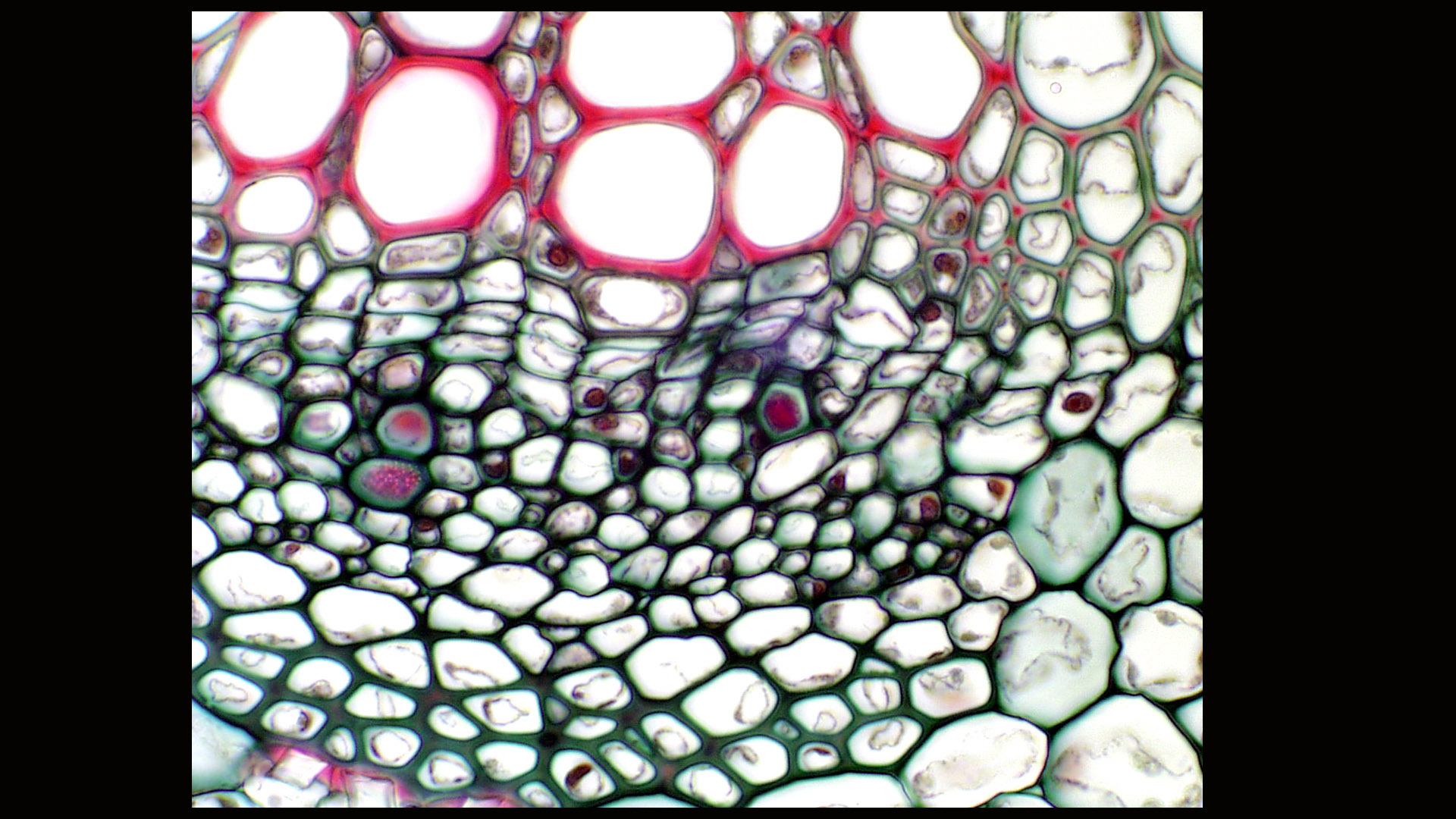
Sieve Element Microscope . a method was designed for in vivo observation of sieve element/companion complexes by using confocal laser scanning. we used confocal scanning laser microscopy and subcellular fluorescent markers in. Although it is now more than a hundred years since hartig (1837,. sieve element, in vascular plants, elongated living cells of the phloem, the nuclei of which have fragmented and disappeared and the transverse end walls of. phloem sieve cells, equivalent to sieve (tube) elements in angiosperms, develop in seedless vascular plants and.
from botit.botany.wisc.edu
sieve element, in vascular plants, elongated living cells of the phloem, the nuclei of which have fragmented and disappeared and the transverse end walls of. a method was designed for in vivo observation of sieve element/companion complexes by using confocal laser scanning. Although it is now more than a hundred years since hartig (1837,. phloem sieve cells, equivalent to sieve (tube) elements in angiosperms, develop in seedless vascular plants and. we used confocal scanning laser microscopy and subcellular fluorescent markers in.
Department of Botany
Sieve Element Microscope phloem sieve cells, equivalent to sieve (tube) elements in angiosperms, develop in seedless vascular plants and. sieve element, in vascular plants, elongated living cells of the phloem, the nuclei of which have fragmented and disappeared and the transverse end walls of. a method was designed for in vivo observation of sieve element/companion complexes by using confocal laser scanning. Although it is now more than a hundred years since hartig (1837,. we used confocal scanning laser microscopy and subcellular fluorescent markers in. phloem sieve cells, equivalent to sieve (tube) elements in angiosperms, develop in seedless vascular plants and.
From search.library.wisc.edu
Sieve plates in a longitudinal section of Cucurbita stem prepared Sieve Element Microscope phloem sieve cells, equivalent to sieve (tube) elements in angiosperms, develop in seedless vascular plants and. we used confocal scanning laser microscopy and subcellular fluorescent markers in. sieve element, in vascular plants, elongated living cells of the phloem, the nuclei of which have fragmented and disappeared and the transverse end walls of. Although it is now more. Sieve Element Microscope.
From www.researchgate.net
Three sieve elementspecific proteins are located predominantly in the Sieve Element Microscope a method was designed for in vivo observation of sieve element/companion complexes by using confocal laser scanning. phloem sieve cells, equivalent to sieve (tube) elements in angiosperms, develop in seedless vascular plants and. Although it is now more than a hundred years since hartig (1837,. sieve element, in vascular plants, elongated living cells of the phloem, the. Sieve Element Microscope.
From www.savemyexams.com
Phloem Sieve Tube Elements CIE A Level Biology Revision Notes 2022 Sieve Element Microscope phloem sieve cells, equivalent to sieve (tube) elements in angiosperms, develop in seedless vascular plants and. sieve element, in vascular plants, elongated living cells of the phloem, the nuclei of which have fragmented and disappeared and the transverse end walls of. Although it is now more than a hundred years since hartig (1837,. we used confocal scanning. Sieve Element Microscope.
From exogmtsyy.blob.core.windows.net
Sieve Element Type at Carl Cook blog Sieve Element Microscope we used confocal scanning laser microscopy and subcellular fluorescent markers in. a method was designed for in vivo observation of sieve element/companion complexes by using confocal laser scanning. Although it is now more than a hundred years since hartig (1837,. phloem sieve cells, equivalent to sieve (tube) elements in angiosperms, develop in seedless vascular plants and. . Sieve Element Microscope.
From www.dreamstime.com
Stem with Sieve Cells Under the Microscope Stock Photo Image of plant Sieve Element Microscope phloem sieve cells, equivalent to sieve (tube) elements in angiosperms, develop in seedless vascular plants and. sieve element, in vascular plants, elongated living cells of the phloem, the nuclei of which have fragmented and disappeared and the transverse end walls of. we used confocal scanning laser microscopy and subcellular fluorescent markers in. a method was designed. Sieve Element Microscope.
From ar.inspiredpencil.com
Sieve Tube Elements Sieve Element Microscope sieve element, in vascular plants, elongated living cells of the phloem, the nuclei of which have fragmented and disappeared and the transverse end walls of. phloem sieve cells, equivalent to sieve (tube) elements in angiosperms, develop in seedless vascular plants and. Although it is now more than a hundred years since hartig (1837,. a method was designed. Sieve Element Microscope.
From www.alamy.com
Sieve tube elements of Bryonia sp. Optical microscope X200 Stock Photo Sieve Element Microscope a method was designed for in vivo observation of sieve element/companion complexes by using confocal laser scanning. phloem sieve cells, equivalent to sieve (tube) elements in angiosperms, develop in seedless vascular plants and. sieve element, in vascular plants, elongated living cells of the phloem, the nuclei of which have fragmented and disappeared and the transverse end walls. Sieve Element Microscope.
From www.researchgate.net
Phloem sieve tube geometry. (A) Schematics of a sieve tube. Adjacent Sieve Element Microscope Although it is now more than a hundred years since hartig (1837,. a method was designed for in vivo observation of sieve element/companion complexes by using confocal laser scanning. sieve element, in vascular plants, elongated living cells of the phloem, the nuclei of which have fragmented and disappeared and the transverse end walls of. we used confocal. Sieve Element Microscope.
From www.istockphoto.com
Stem With Sieve Cells Under The Microscope Stock Photo Download Image Sieve Element Microscope phloem sieve cells, equivalent to sieve (tube) elements in angiosperms, develop in seedless vascular plants and. a method was designed for in vivo observation of sieve element/companion complexes by using confocal laser scanning. sieve element, in vascular plants, elongated living cells of the phloem, the nuclei of which have fragmented and disappeared and the transverse end walls. Sieve Element Microscope.
From www.sciencephoto.com
Phloem sieve plates in plant stem Stock Image B725/0281 Science Sieve Element Microscope we used confocal scanning laser microscopy and subcellular fluorescent markers in. sieve element, in vascular plants, elongated living cells of the phloem, the nuclei of which have fragmented and disappeared and the transverse end walls of. a method was designed for in vivo observation of sieve element/companion complexes by using confocal laser scanning. Although it is now. Sieve Element Microscope.
From www.alamy.com
Sieve tube microscope hires stock photography and images Alamy Sieve Element Microscope Although it is now more than a hundred years since hartig (1837,. a method was designed for in vivo observation of sieve element/companion complexes by using confocal laser scanning. phloem sieve cells, equivalent to sieve (tube) elements in angiosperms, develop in seedless vascular plants and. we used confocal scanning laser microscopy and subcellular fluorescent markers in. . Sieve Element Microscope.
From www.researchgate.net
Sieve tubes (ST) with compound sieve plates (spl), uniseriate and Sieve Element Microscope Although it is now more than a hundred years since hartig (1837,. sieve element, in vascular plants, elongated living cells of the phloem, the nuclei of which have fragmented and disappeared and the transverse end walls of. we used confocal scanning laser microscopy and subcellular fluorescent markers in. a method was designed for in vivo observation of. Sieve Element Microscope.
From mavink.com
Sieve Tube Diagram Sieve Element Microscope we used confocal scanning laser microscopy and subcellular fluorescent markers in. a method was designed for in vivo observation of sieve element/companion complexes by using confocal laser scanning. Although it is now more than a hundred years since hartig (1837,. phloem sieve cells, equivalent to sieve (tube) elements in angiosperms, develop in seedless vascular plants and. . Sieve Element Microscope.
From www.kbg.fpv.ukf.sk
sieve plate, side Sieve Element Microscope sieve element, in vascular plants, elongated living cells of the phloem, the nuclei of which have fragmented and disappeared and the transverse end walls of. phloem sieve cells, equivalent to sieve (tube) elements in angiosperms, develop in seedless vascular plants and. a method was designed for in vivo observation of sieve element/companion complexes by using confocal laser. Sieve Element Microscope.
From propg.ifas.ufl.edu
Cell Types, Phloem Sieve Element Microscope Although it is now more than a hundred years since hartig (1837,. sieve element, in vascular plants, elongated living cells of the phloem, the nuclei of which have fragmented and disappeared and the transverse end walls of. phloem sieve cells, equivalent to sieve (tube) elements in angiosperms, develop in seedless vascular plants and. we used confocal scanning. Sieve Element Microscope.
From www.researchgate.net
(PDF) Identification of sieve elements and companion cell protoplasts Sieve Element Microscope phloem sieve cells, equivalent to sieve (tube) elements in angiosperms, develop in seedless vascular plants and. we used confocal scanning laser microscopy and subcellular fluorescent markers in. sieve element, in vascular plants, elongated living cells of the phloem, the nuclei of which have fragmented and disappeared and the transverse end walls of. Although it is now more. Sieve Element Microscope.
From exogmtsyy.blob.core.windows.net
Sieve Element Type at Carl Cook blog Sieve Element Microscope a method was designed for in vivo observation of sieve element/companion complexes by using confocal laser scanning. phloem sieve cells, equivalent to sieve (tube) elements in angiosperms, develop in seedless vascular plants and. Although it is now more than a hundred years since hartig (1837,. sieve element, in vascular plants, elongated living cells of the phloem, the. Sieve Element Microscope.
From www.pnas.org
Proteomics of isolated sieve tubes from Nicotiana tabacum sieve Sieve Element Microscope Although it is now more than a hundred years since hartig (1837,. sieve element, in vascular plants, elongated living cells of the phloem, the nuclei of which have fragmented and disappeared and the transverse end walls of. a method was designed for in vivo observation of sieve element/companion complexes by using confocal laser scanning. we used confocal. Sieve Element Microscope.
